About
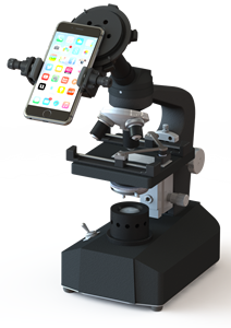
The device, developed by Syntheslide,
that mounts a cell phone onto
a microscope ocular
Syntheslide is a digital pathology start-up company focused on cost effective mobile whole slide image production.
One of our main products includes a universally standardizing portable mechanism that connects your cell phone to a microscope ocular. This is used in conjunction with a mobile application to not only capture images of what the human eye would see through a microscope lens, but to also stitch these individual images into a complete digital pathology slide. Our device, app, and protocol has application beyond whole slide imaging production in a variety of clinical settings.
We have developed a Whole Slide Imaging (WSI) protocol that acquires image data from an externally mounted digital camera/smartphone on a microscope ocular. We developed a universal adapter to standardize camera placement with respect to the microscope ocular. An associated smartphone application sends imaging data for our server to algorithmically post-process into an interactive WSI, accessible through our mobile app and our website. Most pathology labs already implement the use of digital scanning machines or already have some form of digital pathology telemedicine consultation solution in place. Digital slide scanning machines can be many thousands of dollars of expenditures and are not yet a cost effective solution for all labs of the world. Moreover, the cost of software integration of imaging data into hospital EMRs can well surpass the overall cost of hardware. Our protocol is unique in that it leverages existing technology to empower any individual with an internet connection with the capacity to rapidly produce WSIs of diagnostic value at virtually no cost. The WSI data obtained through our protocol is accessible to the user through our application. Users can set sharing settings on WSIs to colleagues and professional contacts.
As with any new protocol for creation of WSI, validation studies are crucial to ensure that diagnostic performance that relies on any novel protocol for digitizing slides is at least equivalent to that of glass slides and light microscopy. The College of American Pathologists has 12 guideline recommendations for pathology laboratories to internally validate any new WSI technology they use in diagnosis. CAP recommends that each institution internally validates any new WSI protocol, so it makes sense for any large gathering of data using our software to be collaborative. We are therefore pursuing validation studies in a diverse range of clinical laboratories, community practices, and hospitals to ensure the validity of the data we obtain.
Sample Output of High Grade Urothelial Carcinoma
Manually-Obtained, Uncalibrated Image. Approximate scanning time: 40 seconds